Phenylmethylsulfonyl Fluoride → Sodium Lauryl Sulfate
Reaction equation

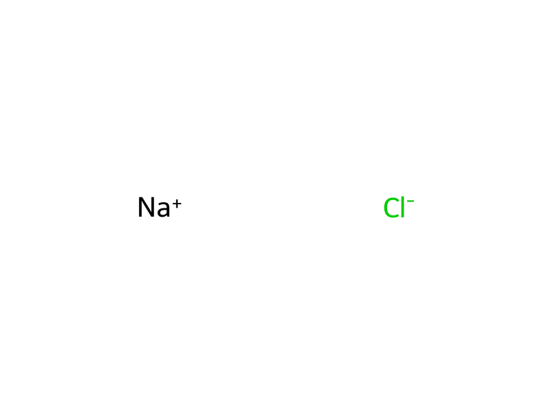
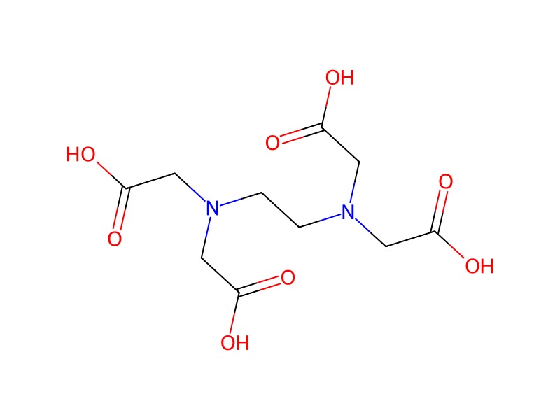
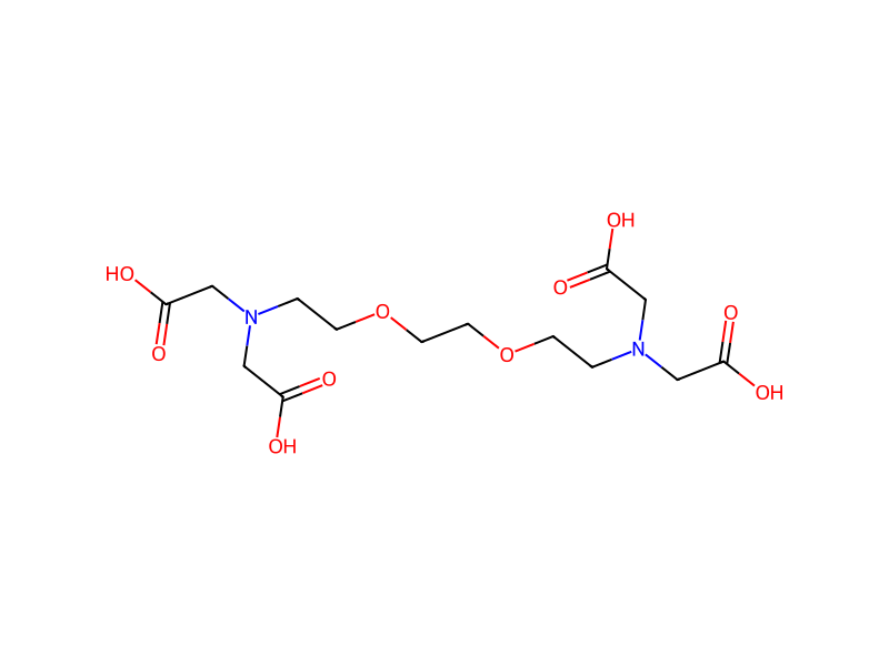
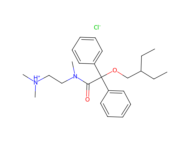
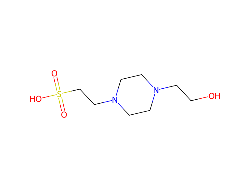
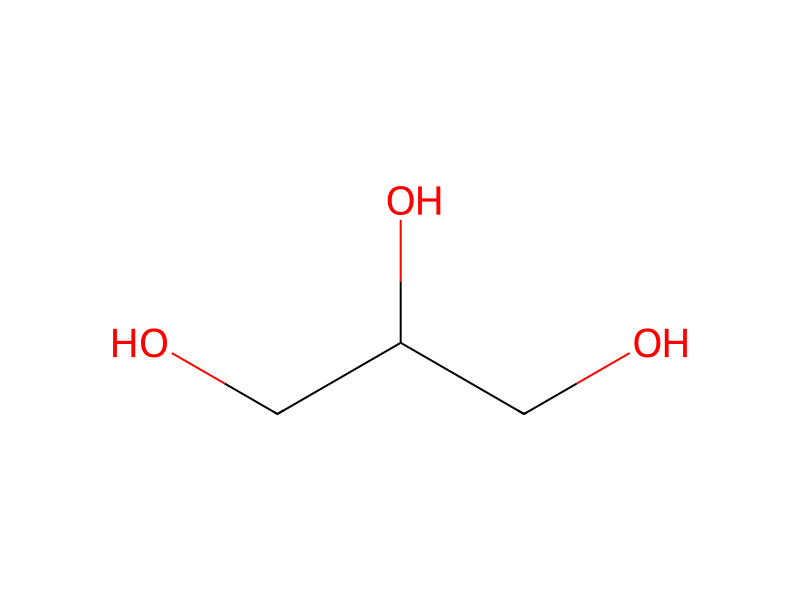
C1N(CCO)CCN(CC[S:12]([OH:15])(=[O:14])=[O:13])C1.[Na+:16].[Cl-].C(N([CH2:40][C:41]([OH:43])=O)CC(O)=O)COCCOCCN(CC(O)=O)CC(O)=O.C(N(CC(O)=O)CC(O)=O)CN(CC(O)=O)CC(O)=O.CCC(CO[C:71](C(N(CC[NH+](C)C)C)=O)([C:78]1[CH:83]=[CH:82][CH:81]=[CH:80][CH:79]=1)[C:72]1C=CC=[CH:74][CH:73]=1)CC.[Cl-].C1C=CC(CS(F)(=O)=O)=CC=1>OCC(CO)O>[CH3:74][CH2:73][CH2:72][CH2:71][CH2:78][CH2:79][CH2:80][CH2:81][CH2:82][CH2:83][CH2:40][CH2:41][O:43][S:12]([O-:15])(=[O:13])=[O:14].[Na+:16]Participants


Conditions
- Temperature
- 96 °C
Procedure
RINm5F cells following different treatments were lysed in a lysis buffer (pH 7.5) consisting of 50 mM HEPES, 150 mM NaCl, 1 EGTA, 1 mM EDTA, 10% glycerol, 1% triton X-100, 1 mM PMSF and a protease inhibitor cocktail (Roche Diagnostics, Mannheim, Germany). The lysate was centrifuged at 800×g for 10 min at 4° C. to remove cell debris and nuclei. The protein concentration of the resulting samples was determined with Bio-Rad protein assay reagent (Bio-Rad, Hercules, Calif.). The samples were then denatured by heating at 96° C. for 3 min in SDS sample buffer and underwent sodium dodecyl sulfate-polyacrylamide gel electrophoresis (SDS-PAGE) and immunoblot analysis. Briefly, 50, 90 or 180 μg proteins were separated in discontinuous gels consisting of a 4% acrylamide stacking gel (pH 6.8) and an 8% acrylamide separating gel (pH 8.8). The separated proteins were then electroblotted to hydrophobic polyvinylidene difluoride membrane (Hybond-P; GE Healthcare, Uppsala, Sweden). The blots were blocked by incubation for 1 h with 5% non-fat milk powder in a washing buffer, containing 50 mM tris(hydroxymethyl)aminomethane, 150 mM NaCl and 0.05% Tween 20 (pH 7.5). They were then incubated overnight at 4° C. with affinity-purified rabbit polyclonal antibodies to β1 integrin (1:500; Millipore, Billerica, Mass.), CaV1.2 (1:200) and CaV1.3 (1:200), respectively, and for 1 h at room temperature with mouse monoclonal antibody to glyceraldehyde-3-phosphate dehydrogenase (GAPDH, 1:4000; Applied Biosystems/Ambion, Austin, Tex.), respectively. After rinsing with the washing buffer, the blots were incubated with the secondary antibodies (either horseradish peroxidase-conjugated goat anti-rabbit IgG or horseradish peroxidase-conjugated goat anti-mouse IgG; 1:50,000; Bio-Rad, Hercules, Calif.) at room temperature for 45 min. The immunoreactive bands were visualized with the ECL plus Western blotting detection system (GE Healthcare, Uppsala, Sweden).
Workup
- customto remove cell debris
- customBriefly, 50, 90 or 180 μg proteins were separated in discontinuous gels
- waitThe blots were blocked by incubation for 1 h with 5% non-fat milk powder
- additionin a washing buffer, containing 50 mM tris(hydroxymethyl)aminomethane, 150 mM NaCl and 0.05% Tween 20 (pH 7.5)
- waitThey were then incubated overnight at 4° C.
- customwith affinity-purified rabbit polyclonal antibodies to β1 integrin (1:500; Millipore, Billerica, Mass.), CaV1.2 (1:200) and CaV1.3 (1:200)
- waitrespectively, and for 1 h at room temperature with mouse monoclonal antibody to glyceraldehyde-3-phosphate dehydrogenase (GAPDH, 1:4000; Applied Biosystems/Ambion, Austin, Tex.)
- washAfter rinsing with the washing buffer, the blots
- waitwere incubated with the secondary antibodies (either horseradish peroxidase-conjugated goat anti-rabbit IgG or horseradish peroxidase-conjugated goat anti-mouse IgG; 1:50,000; Bio-Rad, Hercules, Calif.) at room temperature for 45 min
Sources & evidence
- ORD record
- ord-5c72ae37c3544c1cbdf6f1b7457eea32
- Source dataset
- uspto-grants-2013_10
- Patent number
- US08557513B2
Reaction notes
Loading…