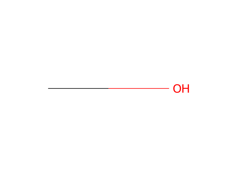
Methanol → Alpha-Cyano-4-Hydroxycinnamic Acid
Reaction equation

C1CCC(NCCCS(O)(=O)=O)CC1.CCN(C1C=CC([C:35]([C:56]2[CH:61]=[CH:60][C:59]([NH:62]C3C=CC(OCC)=CC=3)=[CH:58][CH:57]=2)=[C:36]2C=CC(=[N+](CC3C=CC=C(S([O-])(=O)=O)C=3)CC)C=C2)=CC=1)CC1C=CC=C(S([O-])(=O)=O)C=1.[Na+].C1C=CC(NC2C=CC(N=NC3C=C(S(O)(=O)=O)C=C4C=C(S(O)(=O)=O)C=[C:101]([OH:102])C=34)=C3C=CC=C(S(O)(=O)=O)C=23)=CC=1.CCCCCCCCCCCC[O:127]S([O-])(=O)=O.[Na+].[CH3:133][OH:134]>CO.O.C(O)(=O)C>[OH:134][C:133]1[CH:36]=[CH:35][C:56]([CH:57]=[C:58]([C:59]#[N:62])[C:101]([OH:102])=[O:127])=[CH:61][CH:60]=1Participants


Procedure
For N-terminal sequence analysis and Western blotting, proteins were transferred onto a PVDF membrane (Problott, Applied Biosystems) using a transblot cell (Bio-Rad). The PVDF membrane was wetted in 100% methanol and soaked in transfer buffer (10 mM CAPS, 10% v/v methanol, pH 11.5). Transfer was performed using a potential difference of 60 V for 90 min. For N-terminal sequencing membranes were stained with 0.1% (w/v) Coomassie brilliant blue R250 in methanol/water/acetic acid for 30 sec and destained in 50% v/v methanol. Protein bands were excised and N-terminal sequences determined using a Hewlett Packard 10005A protein sequencer. For peptide mass fingerprinting analysis; Coomassie blue stained protein bands from SDS-PAGE were excised and subjected to in-gel trypsin digestion and subsequent peptide extraction. Protein bands were excised from the Coomassie Blue stained SDS-PAGE gel and gel pieces were washed in 50 mM NH4HCO3/ethanol 1:1, reduced and alkylated with DTT and iodoacetamide, respectively and digested with sequencing grade modified trypsin (Promega) overnight at 37° C. as previously published Mortz et al. (1996). Electrophoresis 17:925-31]. The peptide extract containing 25 mM NH4HCO3 was then analysed by MALDI-TOF MS using an Ultraflex TOF/TOF instrument (Bruker Daltonics) in positive ion and reflectron mode. A saturated solution of 4-hydroxy-α-cyanocinnamic acid (HCCA) was prepared in 97:3 v/v acetone/0.1% v/v aqueous TFA. A thin layer was prepared by pipetting and immediately removing 2 μL, of this solution onto the 600 μm anchorchips of the target plate. Sample (0.5 μL) was deposited on the thin layers with 2.5 μL of 0.1% v/v aqueous TFA, and allowed to adsorb for 5 min, after which the sample solution was removed, and the thin layers washed once with 10 μL of ice-cold 0.1% v/v aqueous TFA for 1 min. Spectra were calibrated by close external calibration using a standard peptide mix. Proteins were identified by peptide mass fingerprinting against the P. gingivalis database (available from www.tigr.org) using an in-house Mascot search engine. Table 2 shows the peptide sequences used to identify the SDS-PAGE separated protein bands of the antigenic complex. The SDS-PAGE of the complex (FIG. 2) is annotated with the designation of the proteins identified by N-terminal sequencing and peptide mass fingerprinting. The complex was found to consist of: Kgpcat, RgpAcat, RgpAA1, KgpA1, RgpAA3, RgpAA2, KgpA2, HagAA1*, HagAA1**, HagAA3 and HagAA2 as well as partially processed Kgp (residues 1 to 700 and residues 136 to 700). A schematic of the processed domains of RgpA, Kgp and HagA are shown in FIG. 3.
Workup
- customfor 90 min
- extractionsubjected to in-gel trypsin digestion and subsequent peptide extraction
- washwere washed in 50 mM NH4HCO3/ethanol 1:1
- custommodified trypsin (Promega) overnight at 37° C. as previously published Mortz et al. (1996)
- extractionThe peptide extract
- additioncontaining 25 mM NH4HCO3
- customA thin layer was prepared by pipetting
- customimmediately removing 2 μL
- customafter which the sample solution was removed
- washthe thin layers washed once with 10 μL of ice-cold 0.1% v/v aqueous TFA for 1 min
- customby close external calibration
- additionmix
- customseparated protein bands of the antigenic complex
Sources & evidence
- ORD record
- ord-75dd7a5bd61d4935a515cbd22e4f85ad
- Source dataset
- uspto-grants-2014_07
- Patent number
- US08765144B2
Reaction notes
Loading…