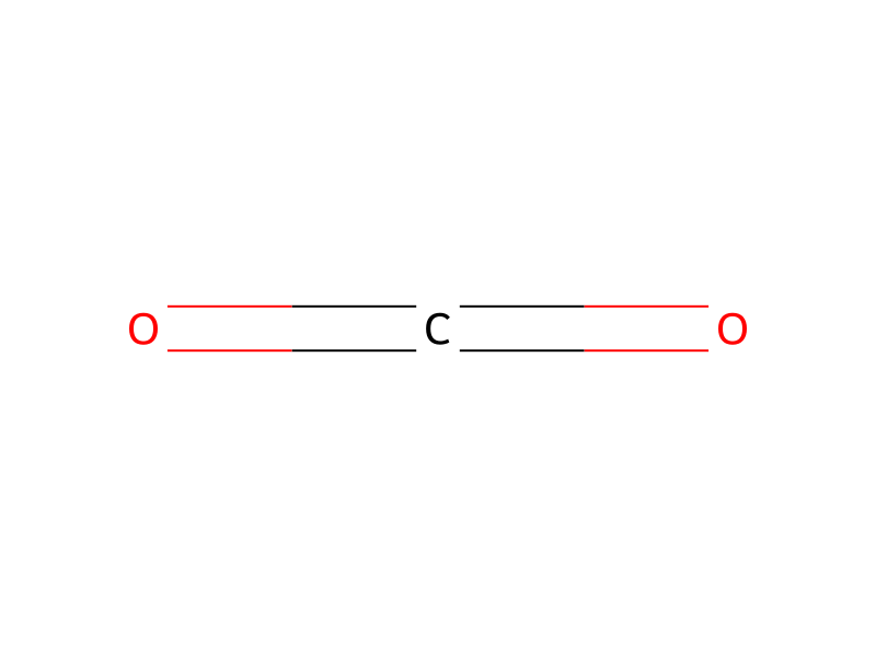
Carbon Dioxide → alpha-Kainic acid
Reaction equation

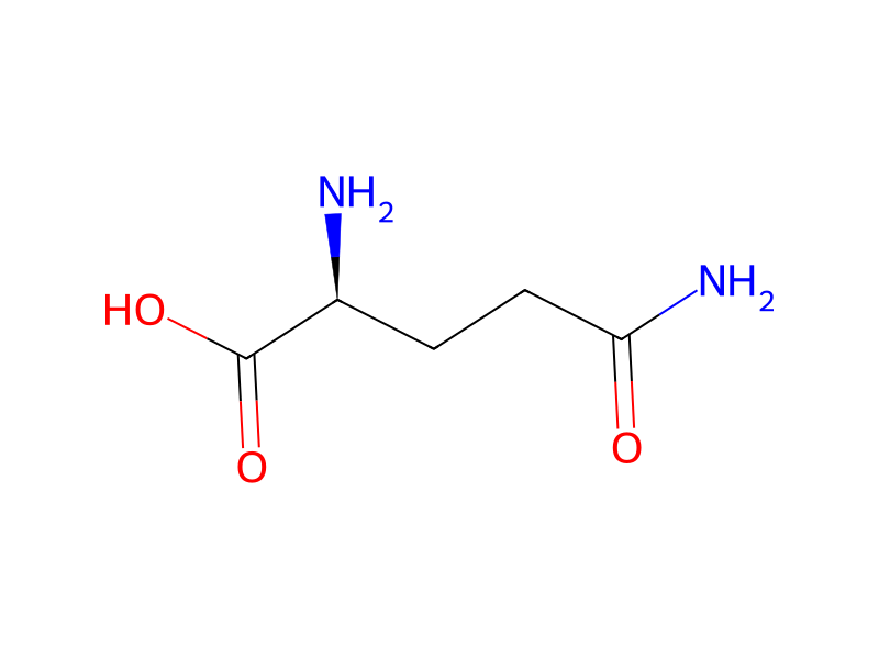
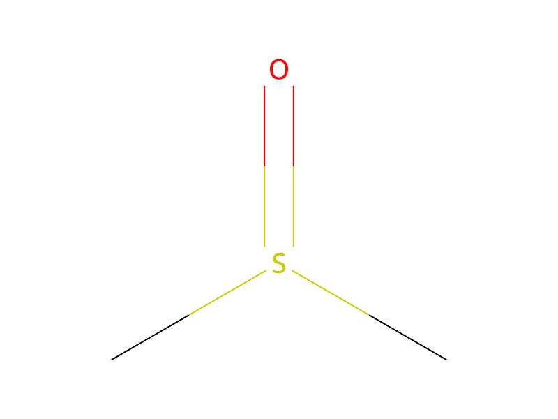
CC1(C)S[C@@H:5]2[C@H:7]([NH:10][C:11]([CH2:13][C:14]3[CH:15]=CC=C[CH:19]=3)=O)[C:8](=[O:9])N2[C@H]1C([O-])=O.[K+].C[C@@H]1[O:30][C@@H:29]([O:31][C@H]2[C@H](O)[C@@H](O)[C@H](NC(N)=N)[C@@H](O)[C@@H]2NC(N)=N)[C@H:28](O[C@@H]2O[C@@H](CO)[C@H](O)[C@@H](O)[C@@H]2NC)[C@@]1(O)C=O.N[C@H](C(O)=O)CCC(=[O:71])N.C(=O)=O>CS(C)=O>[CH3:15][C:14]([C@@H:13]1[C@H:5]([CH2:28][C:29]([OH:31])=[O:30])[C@@H:7]([C:8]([OH:9])=[O:71])[NH:10][CH2:11]1)=[CH2:19]Participants
Reactant 3

Product 1

Reagents, catalysts & solvents 1
Procedure
Cortical neurons were prepared from embryonic day 18 Sprague-Dawley rats. After the cortices had been removed from the brain, isolated cells were plated onto Poly d-Lysine (PDL) (P0899, Sigma)-coated 48 well plates (150687, NUNC) at a density of 2×10⁵ cell per well using Neurobasal Medium (NB) (21103-049, Gibco) with B27 supplement (17504-044, Gibco), penicillin/streptomycin (15140, Gibco) and 1 mM L-glutamine (25030, Gibco). The cell culture was incubated at 37° C. in a humidified atmosphere of 5% CO2 in air. Medium was changed to growth medium (Neurobasal Medium with penicillin/streptomycin and B27 supplement) after 3 hours. Half medium was changed with growth medium every 2-3 days to maintain the cells until day 10 in vitro (DIV 10). Serial dilutions of samples were prepared in growth medium and DMSO was used as vehicle control. For pre-treating assay, half of the medium was removed from wells, and equal amount of diluted samples were replaced. Cells were then incubated for 2 hours at 37° C. in a humidified atmosphere of 5% CO2 in air. Culture was subsequently rinsed with Locke's solution (5 mM potassium chloride, 128 mM sodium chloride, 2.7 mM calcium chloride, 1 mM di-sodium hydrogen orthophosphate, 5 mM HEPES and 10 mM Glucose in Milli-Q water), then incubated with Locke's solution in the presence of glycine (10 μM) for 15 minutes before N-Methyl-D-Aspartic Acid (NMDA) (M-3262, Sigma) treatment. Kainate dissolved in Locke's plus glycine solution was then substituted and incubated for 20 minutes at 37° C. For co-treated kainate assay, serial dilutions of samples were prepared in Locke's solution plus glycine and kainate solution and incubated for 20 minutes at 37° C., 5% CO2. Afterwards, cells were incubated in growth medium for 18-24 hours and detected with Cytotoxicity Detection Kit (1644793, Roche).
Workup
- customCortical neurons were prepared from embryonic day 18 Sprague-Dawley rats
- customAfter the cortices had been removed from the brain
- customisolated cells
- temperatureto maintain the cells until day 10 in vitro (DIV 10)
- additionFor pre-treating assay, half of the medium
- customwas removed from wells, and equal amount of diluted samples
- waitCells were then incubated for 2 hours at 37° C. in a humidified atmosphere of 5% CO2 in air
- washCulture was subsequently rinsed with Locke's solution (5 mM potassium chloride, 128 mM sodium chloride, 2.7 mM calcium chloride, 1 mM di-sodium hydrogen orthophosphate, 5 mM HEPES and 10 mM Glucose in Milli-Q water)
- waitincubated with Locke's solution in the presence of glycine (10 μM) for 15 minutes before N-Methyl-D-Aspartic Acid (NMDA) (M-3262, Sigma) treatment
- dissolutionKainate dissolved in Locke's plus glycine solution
- waitincubated for 20 minutes at 37° C
- additionFor co-treated kainate assay
- customserial dilutions of samples were prepared in Locke's solution plus glycine and kainate solution
- waitincubated for 20 minutes at 37° C.
- waitAfterwards, cells were incubated in growth medium for 18-24 hours
Sources & evidence
- ORD record
- ord-f96d0fc3e33240748433b6c24655cb7b
- Source dataset
- uspto-grants-2015_05
- Patent number
- US09029414B2
Reaction notes
Loading…