Iniparib → Benzamide
HRID 1099494
Reaktionsgleichung

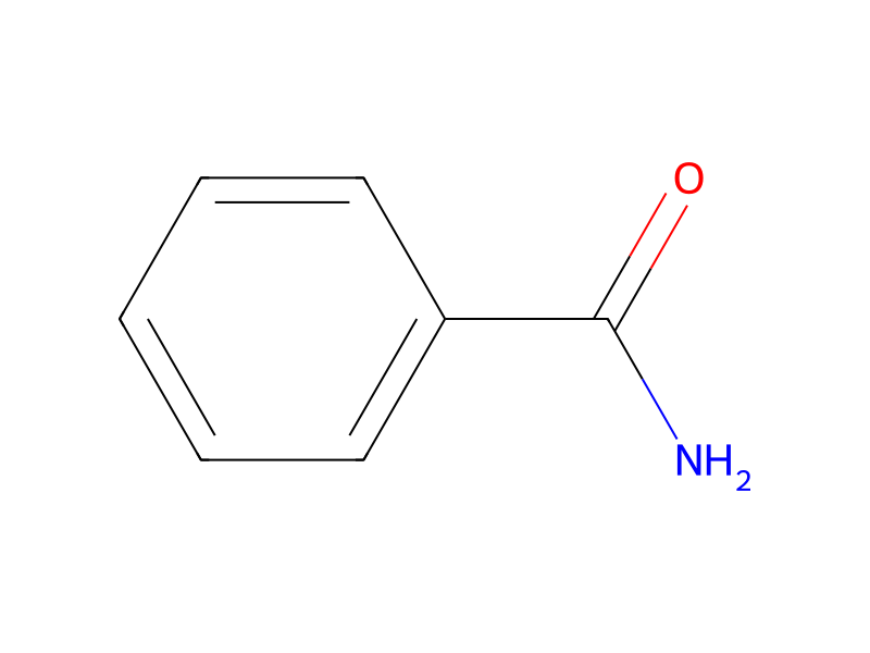
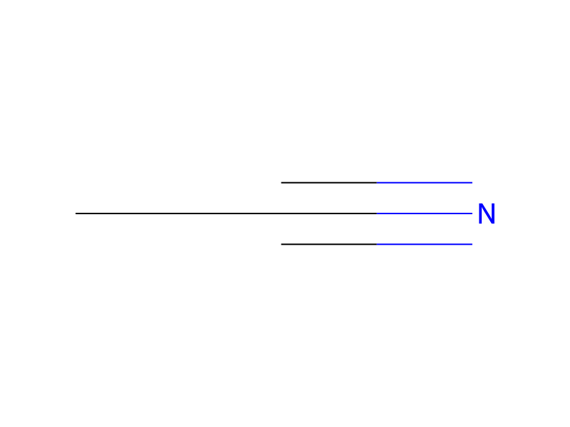
I[C:2]1[CH:10]=[CH:9][C:5]([C:6]([NH2:8])=[O:7])=[CH:4][C:3]=1[N+]([O-])=O>C(#N)C>[C:6]([NH2:8])(=[O:7])[C:5]1[CH:9]=[CH:10][CH:2]=[CH:3][CH:4]=1Komponenten
3
Edukt 1

Produkt 1
Reagenzien, Katalysatoren, Lösungsmittel 1
Versuchsvorschrift
Plasma samples were obtained from dog, rat and mouse studies, in which animals were administered 4-iodo-3-nitrobenzamide (BA). Plasma and tumor samples were prepared for HPLC injection by precipitating plasma (50 μl) with 3× volumes (150 μl) of acetonitrile. Tissue samples were prepared for HPLC injection by adding 1 μl of acetonitrile per mg of tissue, then homogenizing with an electric homogenizer. Following centrifugation, 150 μl of each supernatant was evaporated to dryness, reconstituted in 50 μl of 0.2% formic acid in water and analyzed by chromatography and mass spectrometry techniques (LC/MS/MS conditions). LC/MS/MS conditions are: HPLC (Shimadzu VP System); Mobile Phase: 0.2% formic acid in water (A) and 0.18% formic acid in methanol (B); Column: 1×50 mm Thermo BetaBasic C18 column; Injection volume: 25 μL; Gradient: 0-60% B in 30 minutes; Flow Rate: 100 μL/min; Mass Spectrometer: Applied Biosystems/MDS SCIEX Q-STAR; Interface: IonSpray split at 1/10; Parent Ion Scan: TOF Positive from 200-900 amu; Product Ion Scan: TOF Product Ion from 60-900 amu of most intense Ion in Parent Ion Scan; TOF calibration: Externally calibrated using Renin Substrate. Results of metabolite identification from dog and mouse plasma are depicted in Tables 1-3, as follows: