Sodium Carbonate → Trewsine
반응식

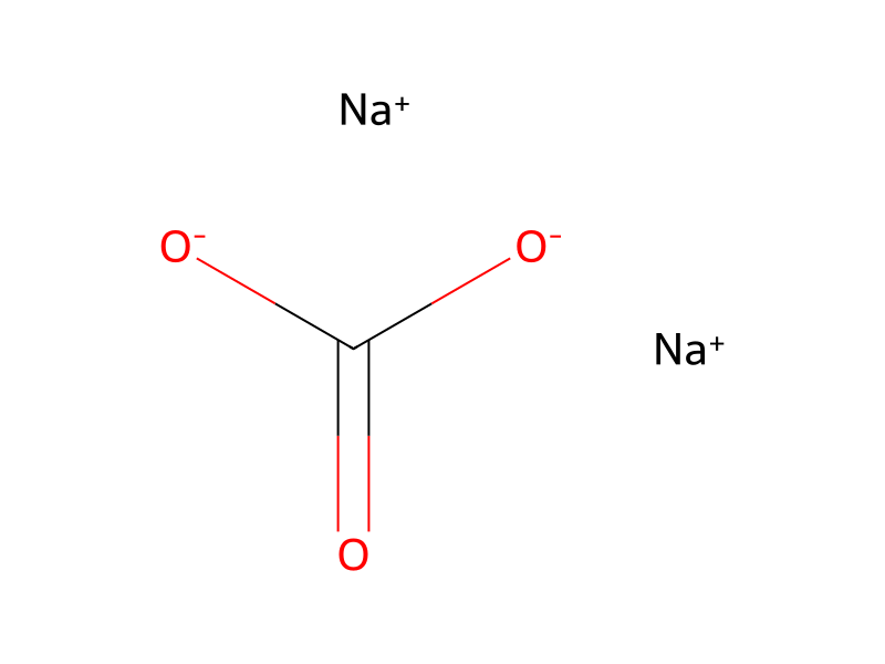
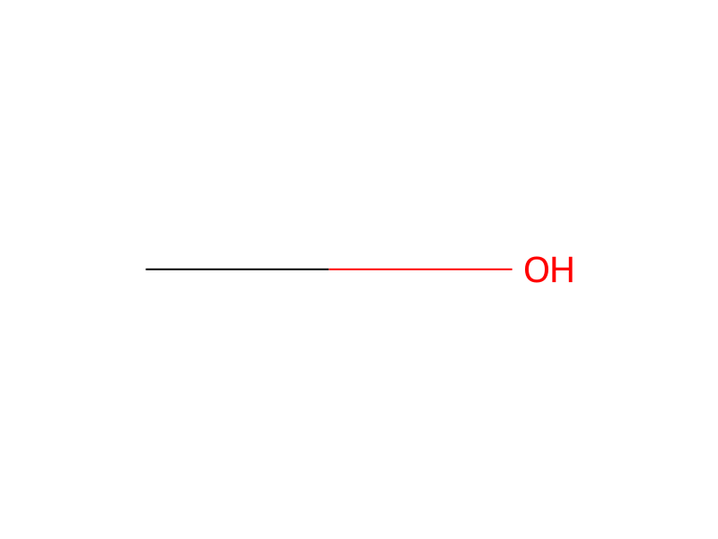
[CH3:1][C@H:2]1[C@@H:27]2[O:28][C@@:26]2([CH3:29])[C@@H:25](OC([C@@H](N(C(C(C)C)=O)C)C)=O)[CH2:24][C:22](=[O:23])[N:21]([CH3:42])[C:14]2=[C:15]([Cl:20])[C:16]([O:18][CH3:19])=[CH:17][C:12](=[CH:13]2)[CH:11]([O:43][CH3:44])[C:10]([CH3:45])=[CH:9][CH:8]=[CH:7][C@@H:6]([O:46][CH3:47])[C@:5]2([OH:52])[NH:48][C:49]([O:51][C@H:3]1[CH2:4]2)=[O:50].C(=O)([O-])[O-].[Na+].[Na+]>CO>[CH3:1][CH:2]1[CH:27]2[O:28][C:26]2([CH3:29])[CH:25]=[CH:24][C:22](=[O:23])[N:21]([CH3:42])[C:14]2=[C:15]([Cl:20])[C:16]([O:18][CH3:19])=[CH:17][C:12](=[CH:13]2)[CH:11]([O:43][CH3:44])[C:10]([CH3:45])=[CH:9][CH:8]=[CH:7][CH:6]([O:46][CH3:47])[C:5]2([OH:52])[NH:48][C:49]([O:51][CH:3]1[CH2:4]2)=[O:50]참가 물질


실험 절차
The structures of the isolated compounds were determined primarily by high resolution mass spectrometry and by proton and carbon 13 NMR using colubrinol and maytansine as standards for comparison. 1H NMR data is given below in Table I and 13C NMR is given in Table II. Comparison of the 1H NMR spectra of trewiasine and colubrinol indicated that these two compounds were closely related, the only significant differences being the presence of an additional methoxyl signal at δ 3.37 in the spectrum of trewiasine and an upfield shift of the C-15 proton singlet from δ 5.48 in colubrinol to δ 4.86 in trewiasine. Referring to Table I, the C-15 proton signals of maytansine (m, δ 3.1-3.0 were replaced by the downfield singlet at δ 4.86 in the spectrum of trewiasine, and as shown in Table II, the C-15 carbon signal of maytansine (t, δ 46.60) was shifted downfield in trewiasine (d, δ 86.70). Thus, trewiasine was determined to have the maytansinoid ring system with a methoxyl at C-15. A mass spectrum gave an apparent M+ -(H2O+HNCO) at m/e 688.0390 (calcd. 688.3126), confirming that trewiasine was C37H52ClN3O11 (MW=749). Both trewiasine and dehydrotrewiasine give a characteristic ion at m/e 515 (C28H34ClNO6) corresponding to an additional loss of the C-3 substituent as consistent with documented M+ -(a+b) type maytansinoid fragmentations. Hydrolysis of trewiasine with sodium carbonate in 50% aqueous methanol at room temperature (see Kupchan, J. Org. Chem. 42, supra) gave trewsine, identified by 1H and 13C NMR, and N-isobutyryl-N-methylalanine, as determined by GC-MS of the methyl ester. This further confirms the structure of trewiasine to be that of 15-methoxymaytanbutine (15-O-methylcolubrinol).
출처 및 근거
- ORD 기록
- ord-6ba216638db044099989e51189788894
- 출처 데이터셋
- uspto-grants-1982_02
- 특허 번호
- US04313946
반응 노트
불러오는 중…