Sodium Chloride → Sodium Lauryl Sulfate
Reaction equation

[Na+:1].[Cl-].C(N(CC(O)=O)CC(O)=O)C[N:5](CC(O)=O)[CH2:6][C:7]([OH:9])=[O:8].CC(CC(C1C=CC(OCCOCCO)=CC=1)(C)C)(C)C.C[C@@H]([C@@H]1[C@@]2(C)[C@@H](O)C[C@@H]3[C@@]4(C)CC[C@@H](O)C[C@H]4CC[C@@]3(C)[C@@H]2CC1)CCC([O-])=O.[Na+].C(N)(=O)C=C.[CH2:79]([OH:86])[C:80]([NH2:85])([CH2:83][OH:84])[CH2:81][OH:82].[CH3:87][CH2:88][CH2:89][CH2:90][CH2:91][CH2:92][CH2:93][CH2:94][CH2:95][CH2:96][CH2:97][CH2:98][O:99][S:100]([O-:103])(=[O:102])=[O:101].[Na+].CN(CCN(C)C)C>>[CH2:79]([OH:86])[C:80]([NH2:85])([CH2:83][OH:84])[CH2:81][OH:82].[NH2:5][CH2:6][C:7]([OH:9])=[O:8].[CH3:87][CH2:88][CH2:89][CH2:90][CH2:91][CH2:92][CH2:93][CH2:94][CH2:95][CH2:96][CH2:97][CH2:98][O:99][S:100]([O-:103])(=[O:102])=[O:101].[Na+:1]Participants
Reactant 8
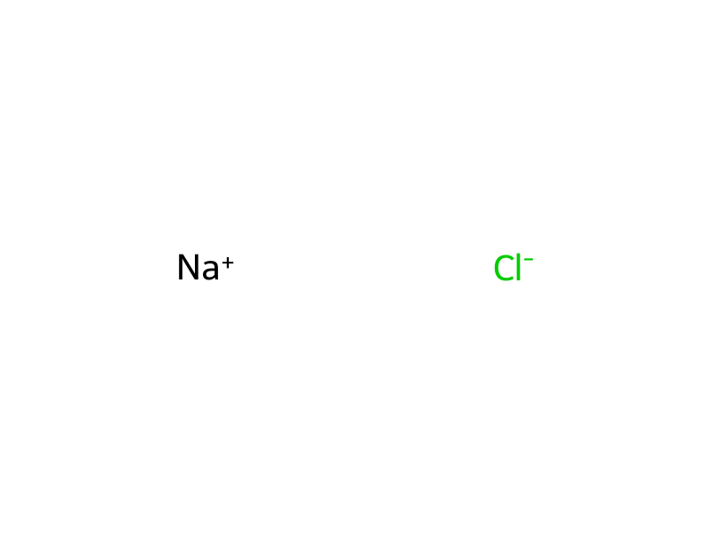 HCID 5234Sodium Chloride
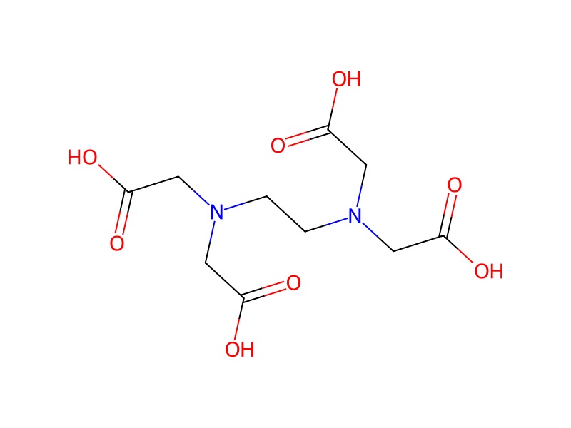 HCID 6049Ethylenediaminetetraacetic Acid2×
 HCID 6503Tris(hydroxymethyl)aminomethane
 HCID 6579Acrylamide
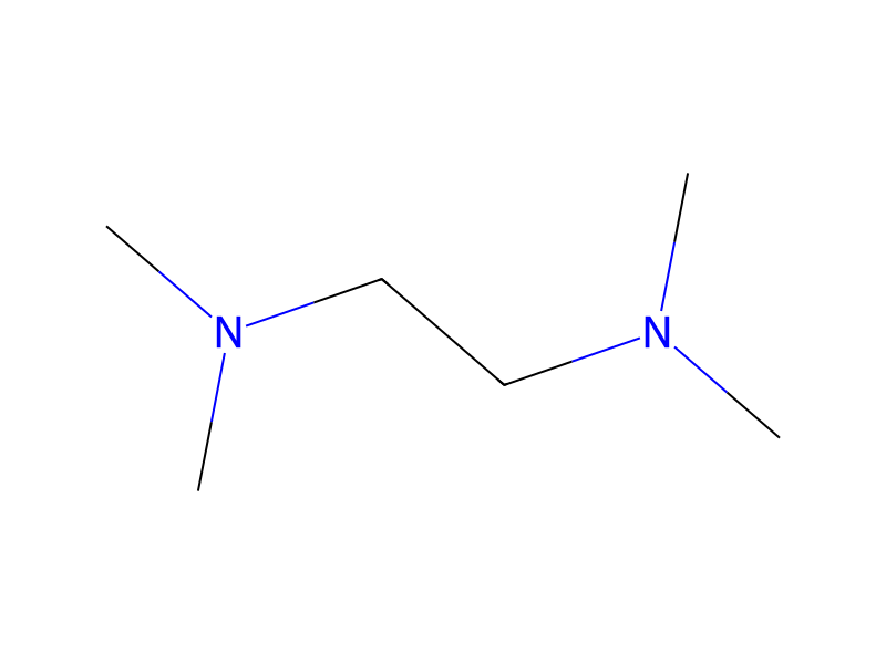 HCID 8037N,N,N',N'-Tetramethylethylenediamine
 HCID 24775Poly(oxy-1,2-ethanediyl), alpha-((1,1,3,3-tetramethylbutyl)phenyl)-omega-hydroxy-, potassium salt (1:1)
 HCID 3423265Sodium Lauryl Sulfate
 HCID 44582061sodium (4R)-4-[(3R,5R,8R,9R,10S,12S,13R,14S,17R)-3,12-dihydroxy-8,10,13-trimethyl-1,2,3,4,5,6,7,9,11,12,14,15,16,17-tetradecahydrocyclopenta[a]phenanthren-17-yl]pentanoate
Product 3
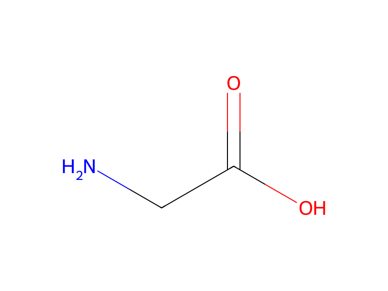
Procedure
Western blotting was performed with the compound of the example 64 of the present invention. A375P cell line was lysed by using RIPA buffer solution (50 mM tris pH 7.4, 150 mM NaCl, 1 mM EDTA, 1% NP-40, 0.25% sodium deoxycholate) containing phosphatase inhibitor (Phosphatase inhibitor cocktail I & II) and protease inhibitor (Complete, Mini, EDTA-free; Roche Applied Science), and centrifuged at 13000 rpm for 10 min. Whole protein was stained with Bio-Rad agent (bio-rad #500-0006) and quantitated by comparing with BSA (positive control). 12% gel was prepared by using 30% acrylamide, 1.5 M Tris (pH 8.8), 10% SDS solution, 10% PAS solution and TEMED. After loading 100 ug of protein per one lane of gel, proteins were isolated by sodiumdodecylsulfate polyacrylamide gel electrophoresis (SDS-PAGE, 250 mM Tris base, 2.5 M Glycine, 0.1% SDS, 140V, electrophoresis for 2 h), and transferred to nitrocellulose filter at 200 mA for 2 h. The results are weakly reacted with an antibody[Rabbit anti-Map Kinase (Zymed #61-7400) and phosphor-p44/42 MAP kinase (Thr202/Tyr204) (Cell signaling #9101)] at 4° C. for a day. Further, it is washed with TBST buffer solution containing 0.05% tween20 at 4 times every 5 minutes. The results are weakly reacted with secondary antibody[anti-rabbit IgG (Amersham #NA934V)] for one hour at room temperature, and washed by the same manner. Protein bands were identified with imaging apparatus (Gelliance). The results is shown in FIG. 1.
Workup
- custom12% gel was prepared
Sources & evidence
- ORD record
- ord-9503b114fcff444ca28fd28e5794819c
- Source dataset
- uspto-grants-2014_06
- Patent number
- US08754209B2
Reaction notes
Loading…