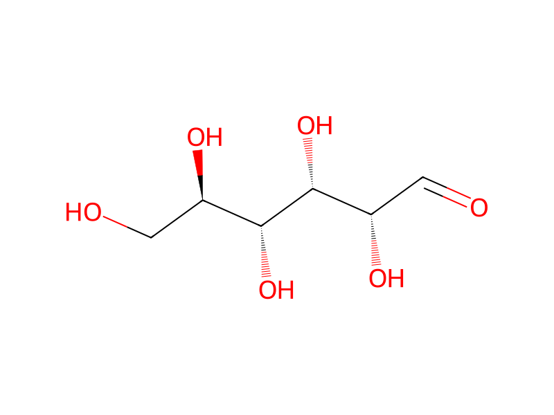
Penicillin G → (2R,3S,4R,5R)-2,3,4,5,6-pentahydroxyhexanal
반응식

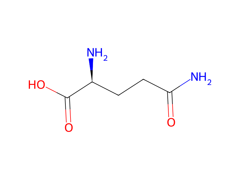
N[C@H](C(O)=O)CCC(=[O:7])N.CC1(C)S[C@@H]2[C@H](NC(CC3C=CC=CC=3)=O)C(=O)N2[C@H]1C(O)=O.C[C@@H]1O[C@@H](O[C@H]2[C@H](O)[C@@H](O)[C@H](NC(N)=N)[C@@H](O)[C@@H]2NC(N)=N)[C@H]([O:58][C@@H:59]2[O:64][C@@H:63]([CH2:65][OH:66])[C@H:62]([OH:67])[C@@H:61]([OH:68])[C@@H:60]2NC)[C@@]1(O)C=O.C[C@@H]1[C@@H](O)[C@@H](C)[C@H](C)OC(=O)C[C@H](O)C[C@H](O)CC[C@@H](O)[C@H](O)C[C@H](O)C[C@@]2(O)O[C@H]([C@H](C(O)=O)[C@@H](O)C2)C[C@@H](O[C@@H]2O[C@H](C)[C@@H](O)[C@H](N)[C@@H]2O)C=CC=CC=CC=CC=CC=CC=C1>>[O:66]=[CH:65][C@@H:63]([C@H:62]([C@@H:61]([C@@H:60]([CH2:59][OH:58])[OH:7])[OH:68])[OH:67])[OH:64]참가 물질
5



실험 절차
Caco-2 cells (5×10⁴) were seeded in a 48-well plate and maintained in culture medium (DMEM with 10% FBS, 1% nonessential amino acids, L-glutamine, penicillin G (100 U/mL), streptomycin (10 μg/mL), and amphotericin B (2.5 μg/mL) in a 37° C. incubator for 10 days for the cells to differentiate. The culture medium was changed once every two days. The cells were then washed with PBS before replenishing with the culture medium containing 5% FBS and various astragalosides at the indicated concentrations (0.01, 0.1 and 1 μM) for 48 hours. The Caco2 cells were washed out of remaining glucose with PBS and replaced in the glucose buffer (80 mM NaCl, 100 mM mannitol, 20 mM Tris-HCl, pH 7.4, 3 mM K2HPO4, 1 mM CaCl2, 1 mg/mL BSA) for 1 hour. Glucose uptake was initiated by replacing the glucose buffer with 0.2 ml of glucose buffer containing 2 μCi/mL of 14C-glucose and unlabeled cold glucose to give a final glucose concentration of 25 mM. Glucose uptake was stopped by removing the glucose buffer and washing with PBS at designated time intervals. The cells were lysed in 0.2 mL of 0.2 N NaOH, and 20 μL of the cell lysate were transferred to the filter-bottomed UniFilter plates (Perkin-Elmer, Wellesley, Mass., USA) and dried in a vacuum oven at 37° C. The bottom of the UniFilter plate was sealed and 25 μL of the counting solution were added into each well. Adhesive plate sealers were used in place of the lids and radioactivity of each sample was counted using the microplate liquid scintillation counter (TopCount, Packard NXT, Packard BioScience Company, Meriden, Conn., USA). The amount of glucose accumulated in the cells was calculated and normalized to protein concentration, and uptake rate was expressed as nanomoles of glucose per minutes per milligram of cell protein (nmol/min/mg). Protein concentration was determined by a standard Bicinchoninic acid (BCA) protein assay. Nonspecific glucose uptake was measured by adding 2 μCi of L-[14C]-glucose and subtracting from each determination to obtain specific glucose uptake.