Sucrose → L-Ascorbic Acid
Reaction equation

C(O)[C@H]1O[C@H](O[C@]2(CO)O[C@H](CO)[C@@H](O)[C@@H]2O)[C@H](O)[C@@H](O)[C@@H]1O.[O:24]=[CH:25][C@@H:26]([C@H:28]([C@@H:30]([C@@H:32]([CH2:34][OH:35])[OH:33])[OH:31])[OH:29])[OH:27]>>[O:24]=[C:25]1[O:31][C@H:30]([C@H:32]([CH2:34][OH:35])[OH:33])[C:28]([OH:29])=[C:26]1[OH:27]Participants
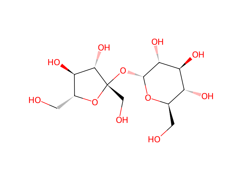
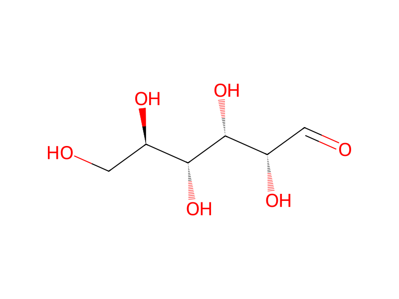
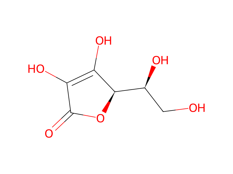
Procedure
Isolation and Culture of Growth Plate Chondrocytes: Six-to eight-week old broiler chickens were obtained commercially, sacrificed by cervical displacement, and the tibiae harvested. Epiphyseal growth plate chondrocytes were obtained from the tibiae as described (Ali et al., 1970, Proc Natl Acad Sci USA 67: 1513-1520). Briefly, cartilage slices were digested in 1% trypsin solution prepared in synthetic cartilage lymph (SCL). SCL contains 2mM Ca2+ and 1.42 mM Pi, in addition to 104.5 mM Na+, 133.5 mM Cl−, 63.5 mM sucrose, 16.5 mM N-tris methyl-2-aminoethane sulfonic acid (TES), 12.7 mM K+, 5.55 mM D-glucose, 1.83 mM HCO3−, 0.57 mM Mg2+, and 0.57 mM SO4−. Cartilage slices were digested for 15 to 20 minutes at 37° C. After digestion, part of the trypsin solution was removed and 10 ml of SCL and 0.5 ml of 0.78% collagenase solution added to the tissue slices. After 30 min of collagenase/trypsin digestion at 34° C., the liquid was removed from the slices and replaced with 0.5 ml of 0.78% collagenase solution plus 15 ml DMEM, with 10% FBS. The collagenase-DMEM solution was removed from the cartilage digestate after 15 hours (overnight) of digestion. The chondrocytes were released from the residual tissues by vortexing for a total of 3 min (1 min each time) in fresh medium. Isolated cells were demineralized in iso-osmotic citrate buffer (8.0 mM citric acid in 80.5 mM sodium citrate, pH 6.0) for 15 min at 34° C. After demineralization, cells were washed with fresh medium, resuspended in 10 ml of DMEM with 10% FBS, and counted using a hemocytometer. Cell viability was determined by trypan blue exclusion. Cells were plated at 3-4×10⁵ cells per 35 mm dish in 2 ml of medium. The cells were initially cultured in DMEM with 10% FBS and 1% penicillin/streptomycin solution. The culture medium was changed twice a week for the duration of the experiments. On day 7, the cell cultures were changed into a 1:1 (v/v) mixture of DMEM with 10% FBS and serum-free HL-1 medium. Ascorbic acid was provided at 50 μg/ml medium after day 3. For treatment of endothelial cell cultures, the concentrated fusarochromanone solution was diluted in methanol to final concentrations of 0.6, 3, 15 mM. On day 7, 2 μl samples of the FC solutions or pure methanol for the control were added to the cultures in 2 ml of media. FIGS. 5A to 5D show there is no appreciable change in the morphology of chondrocytes treated with FC101 for seven days. Note that there is little difference between the control cells (5A) and those treated with FC101 (B:0.6 μM; C:3 μM; D:15 μM FC101).
Workup
- customIsolation and Culture of Growth Plate Chondrocytes: Six-to eight-week old broiler chickens were obtained commercially
- customEpiphyseal growth plate chondrocytes were obtained from the tibiae
- customprepared in synthetic cartilage lymph (SCL)
- customAfter digestion, part of the trypsin solution was removed
- addition10 ml of SCL and 0.5 ml of 0.78% collagenase solution added to the tissue slices
- waitAfter 30 min of collagenase/trypsin digestion at 34° C.
- customthe liquid was removed from the slices
- customThe collagenase-DMEM solution was removed from the cartilage digestate after 15 hours (overnight) of digestion
- customby vortexing for a total of 3 min
- custom1 min
- waitIsolated cells were demineralized in iso-osmotic citrate buffer (8.0 mM citric acid in 80.5 mM sodium citrate, pH 6.0) for 15 min at 34° C
- washAfter demineralization, cells were washed with fresh medium
- waitThe culture medium was changed twice a week for the duration of the experiments
- waitOn day 7
- additionthe cell cultures were changed into a 1:1 (v/v) mixture of DMEM with 10% FBS and serum-free HL-1 medium
Sources & evidence
- ORD record
- ord-b02d9b659ed945d4bc215e4e23891b30
- Source dataset
- uspto-grants-2001_05
- Patent number
- US06225340B1
Reaction notes
Loading…