反応式

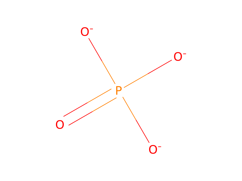
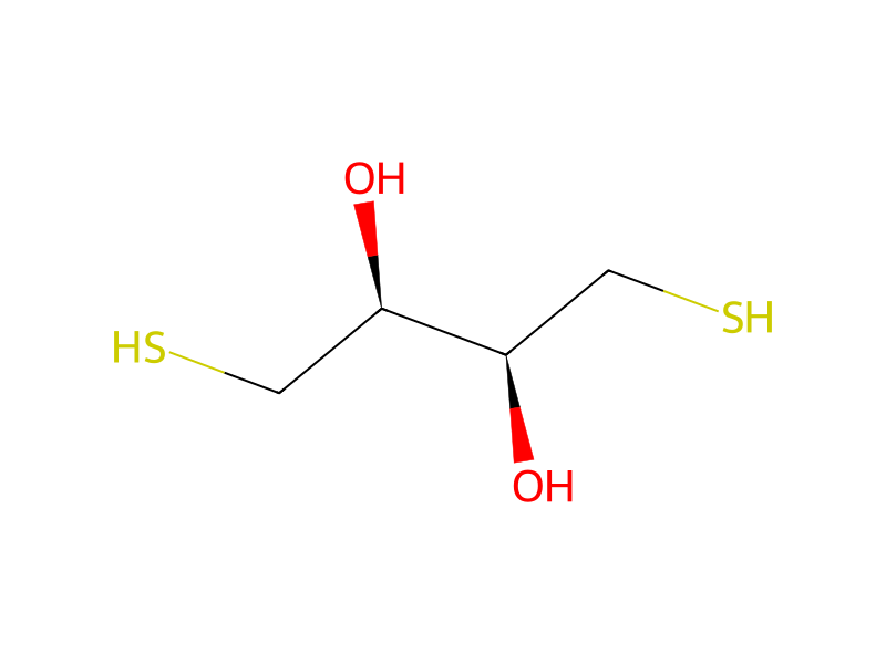
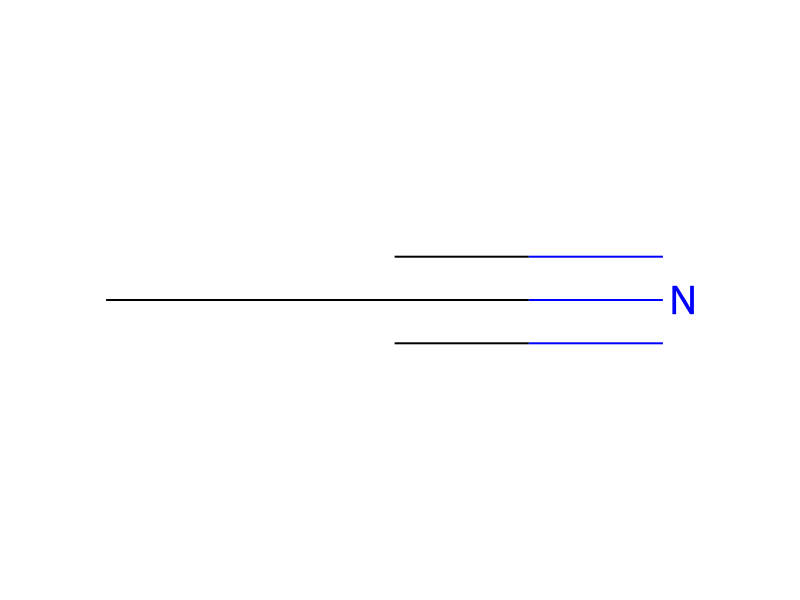
[CH3:1][C@:2]12[C@@H:11]3[CH2:12][CH2:13][C@@:14]4([O:19][C@@H]5O[C@@H](CO)[C@@H](O)[C@H](O)[C@@H]5O[C@@H]5O[C@@H](CO)[C@@H](O)[C@H](O)[C@@H]5O)[C:16]([CH2:18][C@@:10]3([CH2:15]4)[CH2:9][CH2:8][C@@H:7]1[C@@:6]([C:43]([O-:45])=[O:44])([CH3:42])[CH2:5][CH2:4][CH2:3]2)=[CH2:17].[Na+].[K].P([O-])([O-])([O-])=O.SC[C@H]([C@@H](CS)O)O.O[C@H]1O[C@H](CO)[C@@H](O)[C@H](O)[C@H]1O>OP([O-])(O)=O.[K+].CC#N.C(Cl)(Cl)Cl.CO.O>[CH3:1][C@:2]12[C@@H:11]3[CH2:12][CH2:13][C@@:14]4([OH:19])[C:16]([CH2:18][C@@:10]3([CH2:15]4)[CH2:9][CH2:8][C@@H:7]1[C@@:6]([C:43]([OH:45])=[O:44])([CH3:42])[CH2:5][CH2:4][CH2:3]2)=[CH2:17]参加物






実験手順
Steviolbioside, 3-sulfopropyl ester, potassium salt prepared in Part B. was incubated anaerobically for three days at 37° C. with 5 wt% fresh rat cecal contents, at concentrations of 0.25, 0.5, and 1.0 mg/ml in sterile Krebs-Ringer 0.25M phosphate buffer (pH 7.4) containing 0.25 mg/ml dithiothreitol and 0.25 mg/ml α-D-glucose. TLC [silica gel F-254; CHCl3 :CH3OH:H2O (15:10:2)] and HPLC [30 cm C-18 on μ-Bondapak; 15 min linear gradient of 10-40% CH3CN in 0.005M KH2PO4 (pH 3.45); 200 nm] analysis showed all the initial ester (Rf=0.42; tR =14.0 min) to have been consumed within 24 h to yield apparently only the sulfopropyl ester of steviol, (Rf =0.63; tR =16.3 min). No steviol (Rf =0.95; tR =31.3 min) was detected. After 3 days the bacterial cells were sedimented by centrifugation. The sediment was extracted (THF), as was the supernatant, after lyophilization. HPLC analysis for steviol of the two THF extracts showed none to be detectable. With a detection limit of 0.05 g, as little as 0.03 and 0.13 percent degradation to steviol could have been detected for the sediment and supernatant fractions, respectively.
後処理
- customtR =14.0 min) to have been consumed within 24 h
出典と根拠
- ORDレコード
- ord-2348d9a2efe5469ba8a16b193cc98e61
- 出典データセット
- uspto-grants-1984_06
- 特許番号
- US04454290
関連ノート
読み込み中…